Abdominal Anatomy : Abdomen Illustrations Visualisations Of Human Anatomy. Terms in this set (94) what is the abdomen. The diaphragm is its upper boundary. Browse 7,532 abdomen anatomy stock photos and images available, or search for digestive system to find more great stock photos and pictures. To reach this goal and minimize complications, every reproductive surgeon requires a thorough knowledge of pelvic anatomy. The muscle fibers of the transversus abdominis run horizontally, similar to a corset or a weight belt.
The abdominal wall surrounds the abdominal cavity, providing it with flexible coverage and protecting the internal organs from damage. The aorta is the largest blood vessel in the body. The region occupied by the abdomen is called the abdominal cavity, and is enclosed by the abdominal muscles at front and to the sides, and by part of the vertebral column at the back. Terms in this set (94) what is the abdomen. The abdominal aorta enters the abdomen through the diaphragm at the level of the twelfth thoracic vertebre and continues to just below the umbilical area, where it splits into the right and left common iliac arteries.
Understanding Abdominal Divisions Anatomy Snippets Complete Anatomy from s3-us-west-1.amazonaws.com The abdominal wall surrounds the abdominal cavity, providing it with flexible coverage and protecting the internal organs from damage. These two apertures, together with abdominal walls, bound the abdominal cavity. The abdomen is the front part of the abdominal segment of the trunk. The left upper quadrant is the location of the left portion of the liver, the larger portion of the. A hernia will usually cause a distinct bulge where the tissue or organ pushes through the muscle wall. Inferiorly the abdomen is open to the pelvis, communicating through the superior pelvic aperture (pelvic inlet). Abdomen, in human anatomy, the body cavity lying between the chest or thorax above and the pelvis below and from the spine in the back to the wall of abdominal muscles in the front. If you plan to enter a healthcare profession such as nursing, this is something you'll use on the job when performing abdominal assessments (and while documenting).
Knowledge of abdominal wall anatomy is important to ensure safe placement of primary and secondary laparoscopic ports.
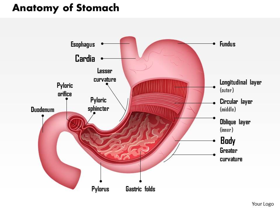
The diaphragm is its upper boundary. To reach this goal and minimize complications, every reproductive surgeon requires a thorough knowledge of pelvic anatomy. The abdominal (peritoneal) cavity is an area that normally only contains a small amount of peritoneal fluid, however can become a potential space for pathology. Knowledge of abdominal wall anatomy is important to ensure safe placement of primary and secondary laparoscopic ports. Stomach is a muscular bag forming the most distensible part of the human digestive system. The area occupied by the abdomen is called the abdominal cavity. We're going to take apart a plastic anatomy model and see what we can find in the abdomen. The abdomen (colloquially called the belly, tummy, midriff or stomach) is the part of the body between the thorax (chest) and pelvis, in humans and in other vertebrates. The component of the urinary system, kidney and the ureter. It is bounded superiorly by the xiphoid process and costal margins, posteriorly by the vertebral column and inferiorly by the pelvic bones and inguinal ligament. The abdomen is the front part of the abdominal segment of the trunk. Terms in this set (94) what is the abdomen. The region occupied by the abdomen is called the abdominal cavity, and is enclosed by the abdominal muscles at front and to the sides, and by part of the vertebral column at the back.
The abdomen is the front part of the abdominal segment of the trunk. Abdominal muscle strains don't cause a bulge or visible lump. Knowledge of abdominal wall anatomy is important to ensure safe placement of primary and secondary laparoscopic ports. The viewer gets to see the abdominal organs just as the surgeon does while he or she is operating o. Abdominal computed tomography (ct) is a type of medical imaging procedure used to diagnose and monitor internal stomach issues, like cancer, bowel obstruction, and abdominal pain.
0514 Anatomy Of Stomach Medical Images For Powerpoint Powerpoint Presentation Designs Slide Ppt Graphics Presentation Template Designs from www.slideteam.net The left upper quadrant is the location of the left portion of the liver, the larger portion of the. The major organs of the abdomen include the small intestine, large intestine, and stomach. The viewer gets to see the abdominal organs just as the surgeon does while he or she is operating o. The aorta is the largest blood vessel in the body. Abdominal muscle strains don't cause a bulge or visible lump. The abdominal (peritoneal) cavity is an area that normally only contains a small amount of peritoneal fluid, however can become a potential space for pathology. The transverse abdominal muscle wraps around the torso from front to back and from the ribs to the pelvis. These two apertures, together with abdominal walls, bound the abdominal cavity.
Anatomy of the abdomen of a woman, anatomy of the abdomen woman, anatomy of woman's left abdomen, anatomy of woman's lower abdomen, human anatomy, anatomy of the.
The muscle fibers of the transversus abdominis run horizontally, similar to a corset or a weight belt. Terms in this set (94) what is the abdomen. The purpose of the abdominal divisions is to describe regional anatomy in the abdomen, and to help clinicians determine which organ and tissues are involved in a disease based on which regions experience pain. Browse 7,532 abdomen anatomy stock photos and images available, or search for digestive system to find more great stock photos and pictures. The abdomen is the part of the body that contains all of the structures between the thorax (chest) and the pelvis, and is separated from the thorax via the diaphragm. One of the easiest ways to tell if your pain is caused by a hernia or pulled stomach muscle is if you have a bulge or not. It is an artery, meaning that it carries blood away from the heart. The main areas of the abdomen include the abdominal cavity, calot's triangle, the peritoneum, the inguinal canal, and hesselbach's triangle. The transverse abdominal muscle wraps around the torso from front to back and from the ribs to the pelvis. Knowledge of abdominal wall anatomy is important to ensure safe placement of primary and secondary laparoscopic ports. The abdominal aorta enters the abdomen through the diaphragm at the level of the twelfth thoracic vertebre and continues to just below the umbilical area, where it splits into the right and left common iliac arteries. It is bounded superiorly by the xiphoid process and costal margins, posteriorly by the vertebral column and inferiorly by the pelvic bones and inguinal ligament. Its superior aperture faces towards the thorax, enclosed by the diaphragm.
This is a laparoscopic tour of abdominal cavity anatomy. Together, these three turn nutrients into usable energy, as well as help dispose of solid waste. This muscle doesn't help move the spine or the pelvis, but it does help with respiration and breathing. Anatomy of the abdomen of a woman, anatomy of the abdomen woman, anatomy of woman's left abdomen, anatomy of woman's lower abdomen, human anatomy, anatomy of the. We're going to take apart a plastic anatomy model and see what we can find in the abdomen.
Abdominal Anatomy Gist Support International from www.gistsupport.org The diaphragm marks the top of the abdomen and the horizontal line at the level of the top of the pelvis marks the bottom. The viewer gets to see the abdominal organs just as the surgeon does while he or she is operating o. The transverse abdominal muscle wraps around the torso from front to back and from the ribs to the pelvis. The main areas of the abdomen include the abdominal cavity, calot's triangle, the peritoneum, the inguinal canal, and hesselbach's triangle. The majority of these organs are encased in a protective membrane termed the peritoneum. Stomach is a muscular bag forming the most distensible part of the human digestive system. The abdomen is the body region found between the thorax and the pelvis. Browse 7,532 abdomen anatomy stock photos and images available, or search for digestive system to find more great stock photos and pictures.
The abdomen is the body region found between the thorax and the pelvis.
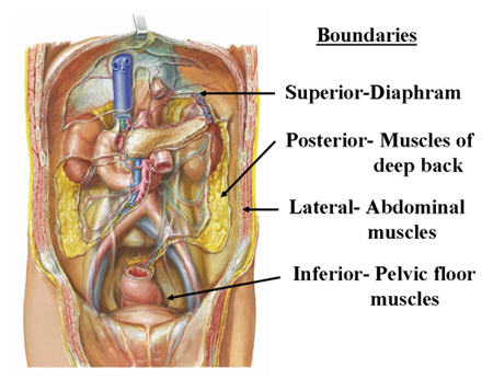
Abdominal computed tomography (ct) is a type of medical imaging procedure used to diagnose and monitor internal stomach issues, like cancer, bowel obstruction, and abdominal pain. This muscle doesn't help move the spine or the pelvis, but it does help with respiration and breathing. Knowledge of abdominal wall anatomy is important to ensure safe placement of primary and secondary laparoscopic ports. The purpose of the abdominal divisions is to describe regional anatomy in the abdomen, and to help clinicians determine which organ and tissues are involved in a disease based on which regions experience pain. We're going to take apart a plastic anatomy model and see what we can find in the abdomen. The aorta is the largest blood vessel in the body. The diaphragm marks the top of the abdomen and the horizontal line at the level of the top of the pelvis marks the bottom. The majority of these organs are encased in a protective membrane termed the peritoneum. The major organs of the abdomen include the small intestine, large intestine, and stomach. The left upper quadrant is the location of the left portion of the liver, the larger portion of the. Browse 7,532 abdomen anatomy stock photos and images available, or search for digestive system to find more great stock photos and pictures. The abdomen (colloquially called the belly, tummy, midriff or stomach) is the part of the body between the thorax (chest) and pelvis, in humans and in other vertebrates. Boundaries of the abdomen (4) anterior abdominal wall (anterolateral) diaphragm (superior)
Share :
Post a Comment
for "Abdominal Anatomy : Abdomen Illustrations Visualisations Of Human Anatomy"


Post a Comment for "Abdominal Anatomy : Abdomen Illustrations Visualisations Of Human Anatomy"